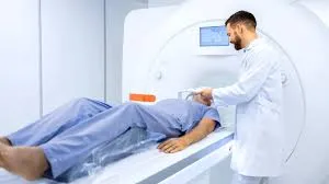
Diagnosing internal organ injuries requires precision and the right tools. Body imaging technologies have transformed how healthcare providers detect and understand injuries. By providing detailed visuals of internal structures, these advanced tools play a pivotal role in effective treatment plans and recovery outcomes. Here is how they support accurate diagnoses:
What Tools Are Typically Used?
Modern medical imaging offers a variety of tools, each designed for specific purposes. These tools allow healthcare professionals to examine internal organs in ways that were not possible decades ago:
- X-ray imaging uses electromagnetic waves to quickly highlight fractures or damage to bones and surrounding organs.
- CT scans, also known as computed tomography, provide cross-sectional images that reveal intricate details of bones, tissues, and blood vessels. They are incredibly reliable for identifying internal bleeding or organ damage.
- MRI imaging, or magnetic resonance imaging, uses magnetic fields and radio waves to generate highly detailed images, especially beneficial for examining soft tissues like the brain or liver.
- Ultrasound imaging, leveraging sound waves, is non-invasive and suitable for real-time imaging of organs such as the heart, kidneys, or even unborn babies.
Each of these techniques delivers insights into the body’s internal state without requiring invasive surgery, which reduces patient risk significantly.
How Do These Techniques Work?
The principles of body imaging are based on technology that visualizes different densities within the body. Each imaging method works differently, depending on what needs to be observed. X-rays pass through soft tissues easily but are absorbed by denser structures like bones, which creates contrast in the image. A CT scan combines multiple X-ray images taken from different angles to produce a three-dimensional view. This is especially useful for injuries involving overlapping structures, such as ribs and lungs.
MRIs measure how water molecules align under strong magnets, providing detailed images of soft tissues like muscles or the brain. Ultrasounds use sound waves that bounce back from structures, allowing real-time visualization of shape and movement. Each method serves specific diagnostic purposes, helping doctors understand the injury clearly before deciding on a treatment plan.
Why Are Imaging Results Reliable?
The reliability of imaging systems comes from their ability to uncover injuries that physical exams might miss. Many symptoms can hide serious complications within the body. Internal bleeding might not be visible from the outside but can be clearly seen on scans. Another benefit is that imaging results are objective. Multiple healthcare providers can examine the same scan and arrive at the same conclusion.
What Benefits Do Patients Gain?
Patients benefit in many ways from imaging technologies. First, non-invasive scans reduce the need for exploratory surgeries, leading to shorter recovery times. Many imaging tests, such as ultrasounds and X-rays, are painless and quick, allowing patients to get results the same day. Additionally, these scans improve diagnostic accuracy for complex conditions.
If someone has unexplained stomach pain, an ultrasound or CT scan can identify the cause, like gallstones or an inflamed appendix. This quick diagnosis helps prevent complications. Seing visual evidence of injuries reassures patients by explaining their symptoms clearly and scientifically, keeping them informed.
Find Body Imaging Options
New technologies like AI-powered imaging software are already changing how efficiently scans are done and making it easier to spot abnormal patterns. Devices with higher resolutions will soon offer more detailed images, helping detect conditions such as cancer or heart disease earlier. Portable imaging tools could also increase access to care in remote areas, removing geographic barriers. To get started, schedule an appointment with a radiologist near you.

